 |
|
Albas |
By Joan Monteith
The female reproductive parts of a rose consist of several parts collectedly known as a pistil. (at right) The outermost part, the stigma, is where pollen is applied. After the pollen germinates, the pollen tube proceeds down the style to the ovary. This picture shows both stigmas and styles. The yellow on the outer edges of the stigmas is pollen.
This is a close-up of one stigma, (at right, below) and the texture
is more noticble. The bumps and protrusions cover the entire surface
of the stigma, although the depth of field of the camera limits what
you can see. Looking at this, it is easier to understand how delicate
the stigmas are, and how fast they can dry out.
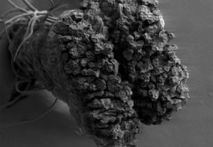
This
is a picture (below, left) of a stigma and style taken using a scanning
electron microscope. The much greater depth of field allows the entire
style/stigma to be seen. When the stigma is ready to receive pollen,
it exudes a clear sticky fluid. This stigma has already produced that
fluid, and that is the wrinkled covering you see over the top of the
stigma.
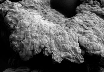
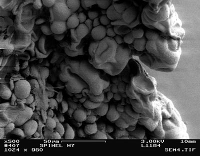
This
is a closeup (at right, below) of the edge of another stigma using
a scanning electron microscope. Again, the sticky fluid has covered
the surface of the stigma, although the angle of the picture gives
a better idea of the surface texture. The faint bumps on the surface
are individual cells. This
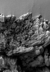
is another scanning electron microscope picture of part (below, left)
of a stigma, perhaps giving a better view of some of the surface texture.
The fluid covering of the stigma is not as thick near the center of
the picture, and the lumps you see are individual cells. This
is an even closer view (at right, below) of part of the stigma in
the previous scanning electron microscope picture.
Original photographs, micrographs and site content (this article) © Joan Monteith 2000.
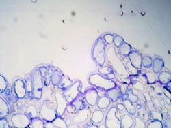
There
have been |
|

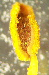 The
anther (left) is the organ at the upper end of a stamen that produces
pollen. The anther in this picture is beginning to discharge the powdery
yellow pollen.
The
anther (left) is the organ at the upper end of a stamen that produces
pollen. The anther in this picture is beginning to discharge the powdery
yellow pollen.
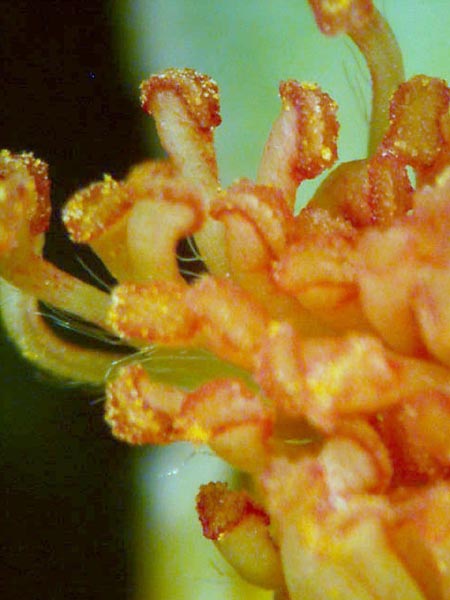
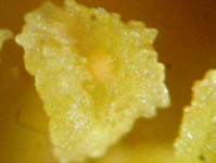
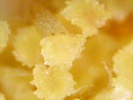 This
is a closer view of some stigmas from another rose. (below, left)
Like stamens, stigmas can come in different colors. The texture of
the stigma--somewhat like a cauliflower--is beginning to show in this
photo.
This
is a closer view of some stigmas from another rose. (below, left)
Like stamens, stigmas can come in different colors. The texture of
the stigma--somewhat like a cauliflower--is beginning to show in this
photo.